Normal shoulder radiograph on X‑ray: 3 real cases
Real, openly licensed radiographs showing normal shoulder radiograph. Study what to look for, then open each case and read it yourself before revealing the answer.
What to look for
- Two orthogonal views: an AP (or true AP / Grashey) plus a scapular Y-view or axial view.
- On a standard AP the humeral head overlaps the glenoid in a smooth ellipse; on the Y-view it sits over the centre of the 'Y'.
- Work round the girdle: glenohumeral joint, proximal humerus, AC joint (inferior borders of acromion and clavicle level), clavicle and scapula.
Quiz yourself on upper limb images
Cases
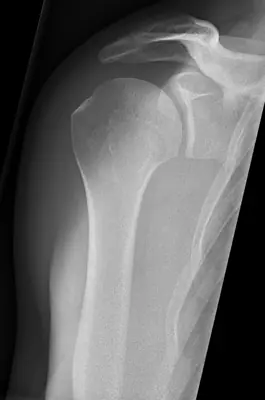 Case #64959082Upper limb · Beginner
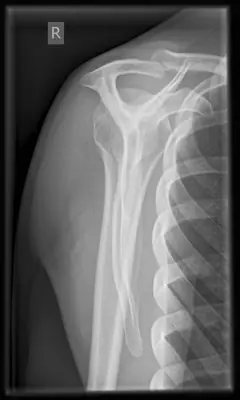 Case #11259059Upper limb · Beginner
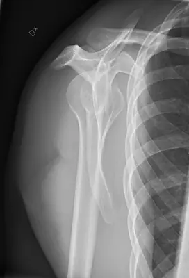 Case #64959081Upper limb · Beginner
Other upper limb diagnoses
- Acromioclavicular joint dislocation
- Calcific tendinitis
- Carpal or carpometacarpal dislocation
- Clavicle fracture
- Distal radius fracture
- Elbow dislocation
- Finger (interphalangeal) dislocation
- Finger (phalangeal) fracture
- Forearm shaft fracture (radius and ulna)
- Galeazzi fracture-dislocation
- Hill-Sachs lesion
- Humeral shaft fracture
- Metacarpal fracture
- Normal elbow radiograph
- Normal hand radiograph
- Normal wrist radiograph
- Proximal humerus fracture
- Radial head fracture
- Rolando fracture
- Scaphoid fracture
- Shoulder (glenohumeral) dislocation
Diagnoses come from each image's documented source. Credits and licences are shown on each case and on the image credits page.