Normal knee radiograph on X‑ray: 6 real cases
Real, openly licensed radiographs showing normal knee radiograph. Study what to look for, then open each case and read it yourself before revealing the answer.
What to look for
- The femur is centred on the tibia on both views and the patella sits at a normal height.
- Both tibial plateaux are smooth, with no step, depression or split, and the joint spaces are similar.
- No effusion or fat-fluid level in the suprapatellar recess on the lateral; a fabella behind the knee is a normal sesamoid.
Quiz yourself on lower limb images
Cases
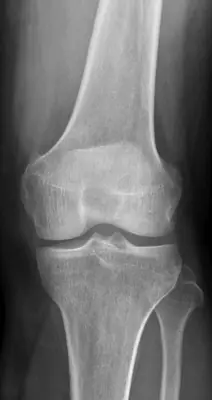 Case #97701195Lower limb · Beginner
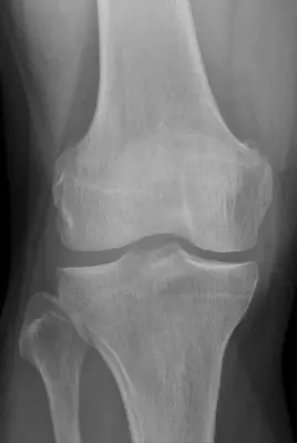 Case #139959249Lower limb · Beginner
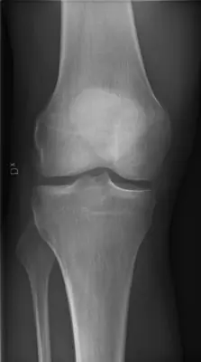 Case #65289734Lower limb · Beginner
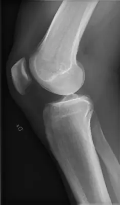 Case #65289732Lower limb · Beginner
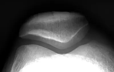 Case #65289731Lower limb · Beginner
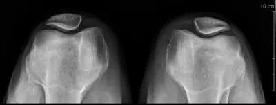 Case #138670513Lower limb · Beginner
Other lower limb diagnoses
- Acetabular fracture
- Ankle fracture (lateral malleolus)
- Bimalleolar ankle fracture
- Calcaneal fracture
- Calcaneal spur
- Femoral shaft fracture
- Fifth metatarsal fracture (Jones / base)
- Hip (proximal femoral) fracture
- Hip dislocation
- Knee joint effusion
- Normal ankle radiograph
- Normal foot radiograph
- Normal pelvis radiograph
- Patellar dislocation
- Patellar fracture
- Pellegrini–Stieda lesion
- Stress fracture
- Tibial (and fibular) shaft fracture
- Tibial plateau fracture
Diagnoses come from each image's documented source. Credits and licences are shown on each case and on the image credits page.