Normal ankle radiograph on X‑ray: 4 real cases
Real, openly licensed radiographs showing normal ankle radiograph. Study what to look for, then open each case and read it yourself before revealing the answer.
What to look for
- A standard series is AP, mortise (about 15-20 degrees of internal rotation) and lateral; the lateral should include the 5th metatarsal base.
- On the mortise view the medial clear space is not widened (about 4 mm or less, and equal to the superior clear space).
- Trace the lateral, medial and posterior malleoli: no cortical break or step.
Quiz yourself on lower limb images
Cases
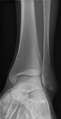 Case #63960357Lower limb · Beginner
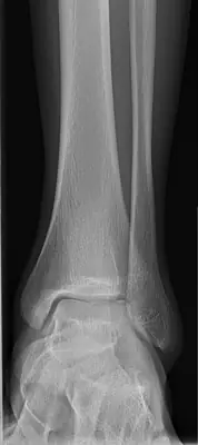 Case #63960358Lower limb · Beginner
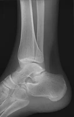 Case #63960360Lower limb · Beginner
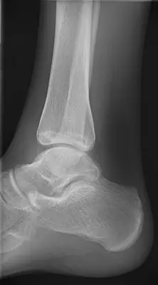 Case #63960356Lower limb · Beginner
Other lower limb diagnoses
- Acetabular fracture
- Ankle fracture (lateral malleolus)
- Bimalleolar ankle fracture
- Calcaneal fracture
- Calcaneal spur
- Femoral shaft fracture
- Fifth metatarsal fracture (Jones / base)
- Hip (proximal femoral) fracture
- Hip dislocation
- Knee joint effusion
- Normal foot radiograph
- Normal knee radiograph
- Normal pelvis radiograph
- Patellar dislocation
- Patellar fracture
- Pellegrini–Stieda lesion
- Stress fracture
- Tibial (and fibular) shaft fracture
- Tibial plateau fracture
Diagnoses come from each image's documented source. Credits and licences are shown on each case and on the image credits page.