Case: Lower limb radiograph #61347912
Lower limb Beginner
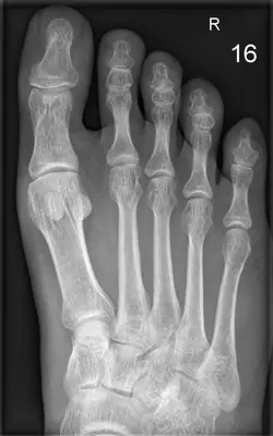
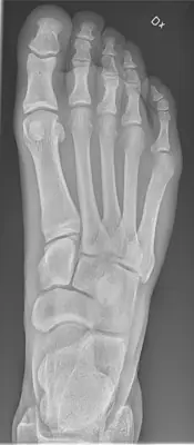
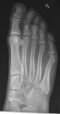
Read this radiograph systematically before revealing the answer. What is the main abnormality?
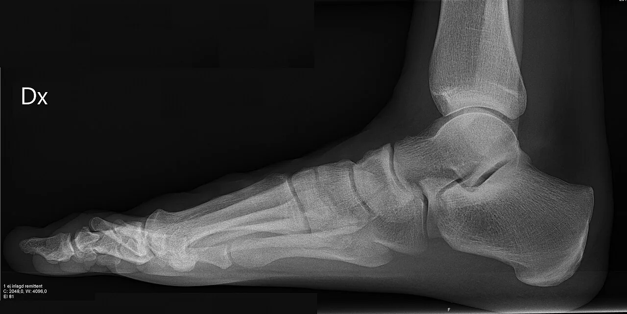
Tip: scroll or pinch to zoom, drag to pan, and try Invert.
Answer: Normal foot radiograph
What to look for in normal foot radiograph
- DP view: the medial border of the 2nd metatarsal base lines up with the medial border of the intermediate cuneiform.
- Oblique view: the 3rd metatarsal lines up with the lateral cuneiform and the 4th with the cuboid.
- Accessory bones and sesamoids are smooth, rounded and fully corticated, unlike acute fracture fragments.
Description by the image author (may be in another language): X-ray ( projectional radiograph ) of a normal right foot of a 31 year old male, by lateral projection. It is a weightbearing image, standing on the imaged foot on a soft material.
Educational case. The diagnosis comes from the image's documented source; the "what to look for" list describes the condition in general and is not a report of this individual image.